
What Is Gynecomastia?
Gynecomastia is abnormal enlargement of male breast tissue caused by a hormonal imbalance—specifically, increased estrogen relative to androgen activity—that stimulates growth of the breast gland itself, not just fat. It affects an estimated 32–55% of men at some point in their lives, making it one of the most common endocrine conditions in males.
The critical distinction is between true gynecomastia (glandular tissue enlargement) and pseudogynecomastia (fatty tissue from weight gain). Only true gynecomastia responds predictably to surgical gland removal; pseudogynecomastia requires liposuction or weight loss. Accurate self-assessment—starting with our online gynecomastia assessment tool—helps you understand which you may have and whether professional evaluation is warranted.
Last medically reviewed by Dr. Sanjog Sharma, MBBS, MS, DNB — August 14, 2026.
Causes and Risk Factors
Gynecomastia occurs when the breast gland proliferates in response to hormonal signals. The main triggers are:
Physiologic gynecomastia — pubertal (age 12–18, affecting ~60–70% of adolescents transiently), neonatal (from maternal estrogen in utero), or age-related (declining testosterone in men over 50).
Medication-induced — antiandrogens like finasteride (used for hair loss) and spironolactone, aromatase inhibitors (which raise estrogen), anabolic steroids and their rebound effect, opioids, and some antipsychotics and antidepressants.
Endocrine and metabolic — hyperthyroidism, hypogonadism, adrenal or pituitary tumors, obesity (adipose tissue converts androgens to estrogen), and liver disease (impaired estrogen metabolism).
Idiopathic — true gynecomastia with no identifiable cause, accounting for ~25% of adult cases.
In my practice at Cocoona Centre for Aesthetic Transformation in Dubai, a significant proportion of male patients presenting for gynecomastia assessment are either taking finasteride for androgenetic alopecia or have a history of anabolic steroid use—often compounded by concurrent weight gain. This dual-trigger scenario is common in fitness-conscious men and athletes.
Self-Assessment: Distinguishing True Gynecomastia from Pseudogynecomastia
Accurate self-assessment begins with understanding what you're looking and feeling for:
 True gynecomastia: firm glandular tissue, often concentrated directly behind and around the nipple-areolar complex (NAC), creating a raised mound.
Clinical Features of True Gynecomastia
- Location and shape: Tissue directly behind and around the nipple-areolar complex (NAC), creating a raised, dome-like mound that projects forward from the chest wall.
- Texture: Firm to rubbery when palpated, not soft like body fat. Glandular tissue feels distinctly different from surrounding adipose tissue.
- Persistence despite weight loss: If you have maintained a stable, healthy BMI or have lost weight and the breast enlargement remains, true gynecomastia is more likely.
- Nipple sensitivity: Mild tenderness or heightened sensitivity to touch is common with glandular tissue.
- Unilateral or bilateral presentation: Can affect one breast (80% of cases) or both (20%).
Clinical Features of Pseudogynecomastia
- Location and shape: Diffuse fullness across the entire chest wall, following the contours of general body fat distribution similar to chest adiposity.
- Texture: Soft, compressible, indistinguishable from surrounding body fat; lacks the firmness of glandular tissue.
- Responds to weight loss or exercise: Decreases significantly when you lose weight or build lean chest muscle (pectoralis major hypertrophy can reduce apparent fullness).
- No nipple involvement: The NAC is flat or even inverted if overall chest fat is prominent; no distinct glandular mound behind it.
The Pinch Test (Home Assessment)
Stand shirtless in front of a mirror:
- Locate the tissue: Use your hand to gently compress your chest directly behind your nipple.
- Pinch test: Gently pinch the tissue between thumb and forefinger for 2–3 seconds.
- Assess texture:
- If you feel distinct, firm tissue (like a disc or nodule) approximately 0.5–2 cm in diameter and separate from surrounding fat, true glandular tissue is likely present.
- If you pinch only soft, compressible fat with no firm glandular component, pseudogynecomastia is more likely.
This home assessment is informative but not diagnostic—it's a useful starting point. Take our online assessment to systematically evaluate your symptoms, and use your results to guide your decision about whether to seek professional consultation.
Age and Presentation Timeline
Pubertal Gynecomastia (Age 12–18)
Accounts for 60–70% of all cases in adolescents. Typically appears around Tanner stage 3–4 (early-to-mid puberty), driven by increased circulating estrogen and relative androgen insufficiency during the early phase of pubertal development.
Natural course: 75% resolve spontaneously within 1–3 years by age 20–21. If gynecomastia persists beyond age 18 or causes significant psychological distress (body image concern, school avoidance, social withdrawal, depression), surgical evaluation is warranted even during teenage years.
Adult-Onset Gynecomastia (Age 25+)
Likely to be persistent without intervention. Common triggers include:
- Recent medication initiation (finasteride, spironolactone, antipsychotics, certain antidepressants).
- Lifestyle changes (significant weight gain, increased alcohol consumption, cannabis use).
- Endocrine shifts (primary hypogonadism, thyroid disease, early andropause or age-related testosterone decline).
- Underlying medical conditions diagnosed incidentally during workup.
Unlike pubertal cases, adult gynecomastia does not typically resolve without treating the underlying cause or surgically removing the glandular tissue.
Diagnosis: What Your Surgeon Will Do
If your self-assessment—or our online gynecomastia assessment tool—suggests possible gynecomastia, a plastic surgeon will:
- Take a detailed history: Onset and progression of enlargement, associated symptoms (pain, discharge from nipple), medication review (including supplements and over-the-counter drugs), family history, alcohol and recreational drug use, recent weight changes.
- Perform a clinical examination: Measure gland size and consistency, assess for firmness vs. laxity, evaluate symmetry between breasts, check for nipple discharge or skin changes, and assess overall chest wall and skin quality.
- Order imaging (if indicated): Chest ultrasound is standard to confirm glandular tissue presence, measure gland volume, assess tissue composition, and exclude breast masses or other pathology. Mammography or MRI is rarely needed unless there are red-flag symptoms (spontaneous discharge, skin dimpling, firm nodules).
- Consider endocrine screening: If gynecomastia is marked, rapidly progressive, recent-onset, or bilateral, blood work (total and free testosterone, estradiol, prolactin, LH, thyroid function, liver function) may identify an underlying endocrine or systemic cause requiring treatment.
At Emirates Hospital in Jumeirah, Dubai, our standard protocol is to perform clinical assessment and chest ultrasound on the first consultation. This combination identifies gland size, tissue composition (true vs. pseudo), and any risk factors—and allows us to counsel on surgical options and recovery at that first visit.
Treatment Options
Conservative Management
Appropriate if:
- Gynecomastia is mild (gland < 2–3 cm) and asymptomatic.
- Pubertal gynecomastia (within the first 12 months of onset; spontaneous resolution is likely).
- The underlying cause is identifiable and reversible (e.g., medication-induced; stopping or switching the offending drug may resolve gynecomastia over 3–6 months).
- The patient is motivated by lifestyle change (weight loss for pseudogynecomastia, alcohol or drug cessation).
- No significant psychological distress.
Medication: Selective estrogen receptor modulators (SERMs) like tamoxifen (10–20 mg daily for 3–6 months) or raloxifene have modest evidence for slowing or arresting glandular growth, particularly if started within 1 year of onset. Success rates for arrest are 60–80%; however, complete reversal of established tissue is rare. Once glandular tissue has fibrosified (> 12 months old), medication is largely ineffective.
Surgical Treatment
Indicated when:
- Gynecomastia is moderate to severe (gland > 2–3 cm) or causing functional limitations (difficulty exercising, clothing restrictions).
- Gynecomastia is causing significant psychological distress (body image concerns, social anxiety, depression).
- Conservative measures have failed or are inappropriate.
- The patient desires definitive, lasting correction.
 Surgical correction via excision and liposuction achieves natural-appearing, lasting results when performed by an experienced plastic surgeon with attention to detail.
Surgical options:
-
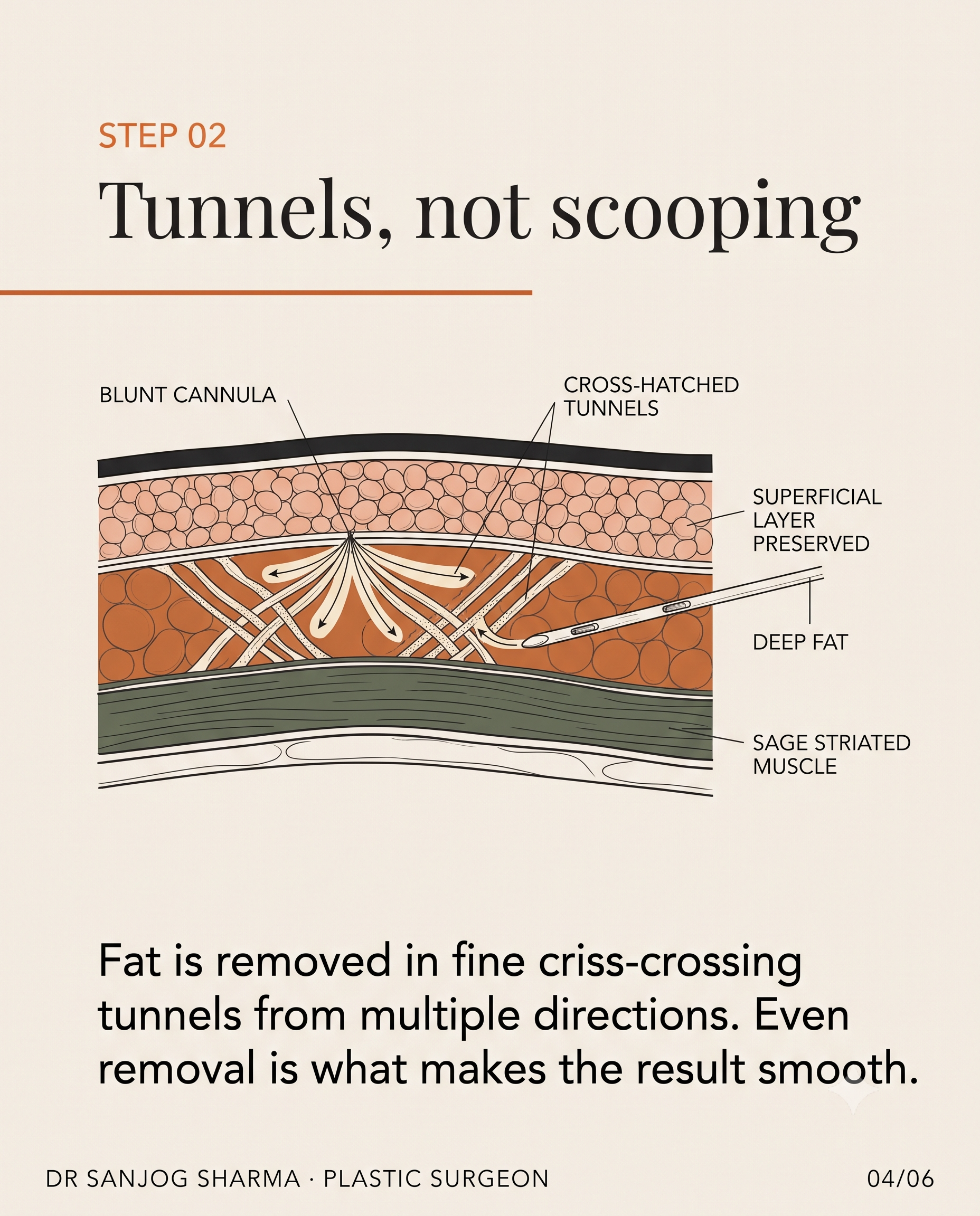
Liposuction alone (for mild gynecomastia with a significant fatty component): Removes fat via small cannulae; suitable when glandular tissue is minimal or absent. Results are excellent for pseudogynecomastia; inadequate for true gynecomastia without gland excision.
-
Excision through a periareolar incision (for moderate gynecomastia with true glandular tissue): Surgeon makes a small curved incision at the edge of the areola, directly excises glandular tissue under direct visualization, and removes excess skin if needed. Leaves minimal, well-camouflaged scarring.
-
Combined liposuction + excision (for gynecomastia with both gland and fat excess): Most common and versatile approach. Liposuction first to remove fat and create better visualization, then excision of glandular tissue under direct control. Allows maximum chest contour refinement and minimal skin trauma.
-
Free nipple graft (for severe gynecomastia with significant skin laxity and NAC repositioning needed): Rarely used; indicated only when the nipple-areolar complex must be repositioned significantly due to severe laxity or very large gland removal. Most gynecomastia cases do not require this.
Surgical goals: Complete removal of all glandular tissue (to prevent recurrence), achievement of chest symmetry, preservation of natural nipple-areolar complex appearance, and minimal, well-hidden scarring.
At Cocoona in Dubai, the vast majority of gynecomastia corrections are performed under local anesthesia with intravenous sedation via a small periareolar excision combined with targeted liposuction, with same-day or next-day discharge and return to light activity within 1–2 weeks.
Recovery Timeline
| Phase | Timeline | What to Expect |
|---|---|---|
| Immediate | Days 1–3 | Surgical dressing in place; mild to moderate pain controlled with prescribed analgesia; compression garment worn continuously; drainage (if placed) monitored. |
| Early | Weeks 1–2 | Dressing removed by day 3–5; compression garment worn 12–18 hours daily; light walking and gentle arm movement encouraged; avoid strenuous chest exercises and heavy lifting. |
| Intermediate | Weeks 3–6 | Drain (if used) removed around day 7–10; gradual return to upper-body activity; light cardio allowed; no heavy pushing, pulling, or contact sports; swelling peaks around week 2–3, then gradually subsides. |
| Late | Weeks 7–12 | Full return to gym, heavy lifting, and contact sports; residual swelling continues to settle; post-operative firmness and sensitivity gradually normalize; scar begins to fade. |
| Final | Months 3–6 | Scar maturation continues; final chest contour clearly visible by 3 months; minor residual swelling or firmness may persist up to 6 months; 95% of patients report satisfaction by 3 months. |
Cost
Gynecomastia surgery in Dubai typically ranges from AED 15,000–35,000 depending on:
- Extent and grade of gynecomastia (mild vs. severe).
- Volume of liposuction required.
- Facility, anesthesia, and operating-room costs.
- Surgeon's experience and credentials.
In Bengaluru at Aesthetica Veda Clinic, the same procedure ranges from INR 1,50,000–3,50,000, offering exceptional value for international and NRI patients.
These are indicative ranges; exact cost is determined after in-person or virtual consultation and ultrasound assessment. Important note: Gynecomastia surgery is typically not covered by insurance in most jurisdictions, as it is classified as cosmetic. Only if gynecomastia arises from a diagnosed medical condition requiring surgical correction (e.g., severe hypogonadism, adrenal pathology) may insurance review coverage—this is rare.
Gynecomastia in Indian Expats and NRI Patients
A significant proportion of my Bengaluru patients are Indian expatriates returning from the UAE, UK, or Australia for surgery. Gynecomastia is highly treatable in both Dubai and Bengaluru, and the choice depends on:
- Timing: Can you allocate 1–2 weeks for surgery and initial recovery?
- Follow-up: Do you prefer to recover near home, or can you return after 3–5 days with remote follow-up via video consultations?
- Cost: Bangalore offers exceptional value (INR 1.5–3.5 lakh) compared to Western centres; Dubai is higher but remains competitive globally.
- Surgeon continuity: Many expats prefer working with the same surgeon for any future needs.
Both Cocoona in Dubai and Aesthetica Veda Clinic in Bengaluru accommodate international and NRI patients; consultations can be conducted online, and pre-operative assessment via local ultrasound can often be arranged before travel, streamlining the process.
Gynecomastia as Part of Broader Body Contouring
For patients combining gynecomastia correction with other body contouring goals—such as liposuction of the abdomen, waist refinement, or post-weight-loss body shaping—a comprehensive surgical plan optimizes results and recovery. Discuss all goals with your surgeon to prioritize procedures and plan staging if multiple surgeries are considered.
Key Points
- Gynecomastia is true glandular breast enlargement from hormonal imbalance, distinct from pseudogynecomastia (fatty enlargement).
- Self-assessment via the pinch test and our online assessment tool helps determine the likelihood of glandular tissue; professional evaluation is essential for diagnosis and treatment planning.
- Pubertal gynecomastia resolves spontaneously in 75% of cases; adult-onset gynecomastia typically requires intervention if bothersome.
- Medication review is critical—finasteride, spironolactone, anabolic steroids, and certain psychiatric medications are common culprits.
- Mild gynecomastia may benefit from conservative management (medication, observation); moderate-to-severe cases are best treated surgically.
- Surgical correction via periareolar excision and liposuction is safe, highly effective, and has > 95% patient satisfaction with < 5% recurrence.
- Recovery to full activity is 6–8 weeks; final contour is fully apparent by 3 months.
- Ready to assess your symptoms? Start with our online gynecomastia assessment tool, then book a consultation with Dr. Sharma at Cocoona Centre for Aesthetic Transformation in Dubai or Aesthetica Veda Clinic in Bengaluru.
References
-
Braunstein GD. Gynecomastia. N Engl J Med. 2007;357:1229–1237. https://doi.org/10.1056/NEJMcp072560
-
Narula HS, Carlson HE. Gynecomastia—pathophysiology, diagnosis, and treatment. Nat Rev Endocrinol. 2014;10:684–698. https://doi.org/10.1038/nrendo.2014.166
-
Johnson RE, Murad MH. Gynecomastia: pathophysiology, evaluation, and management. Mayo Clin Proc. 2009;84(11):1010–1015. https://doi.org/10.4065/84.11.1010
-
Rohrich RJ, Blattner P, Walters J. Gynecomastia: current management. Plast Reconstr Surg. 2015;136(3):430e–442e. https://doi.org/10.1097/PRS.0000000000001539
-
Cantu DE, Amenata ME, Reish RG. Gynecomastia and pseudogynecomastia: review of the literature and functional anatomy of the male chest. Clin Anat. 2018;31(2):202–207. https://doi.org/10.1002/ca.23026
-
Eggener SE, Yossepowitch O, Plas E, et al. Gynecomastia. J Urol. 2014;191(6):1553–1560. https://doi.org/10.1016/j.juro.2014.01.090